AI in Histotechnology: Introduction
A plain explanation of what artificial intelligence is, and where it already sits in the histology laboratory.

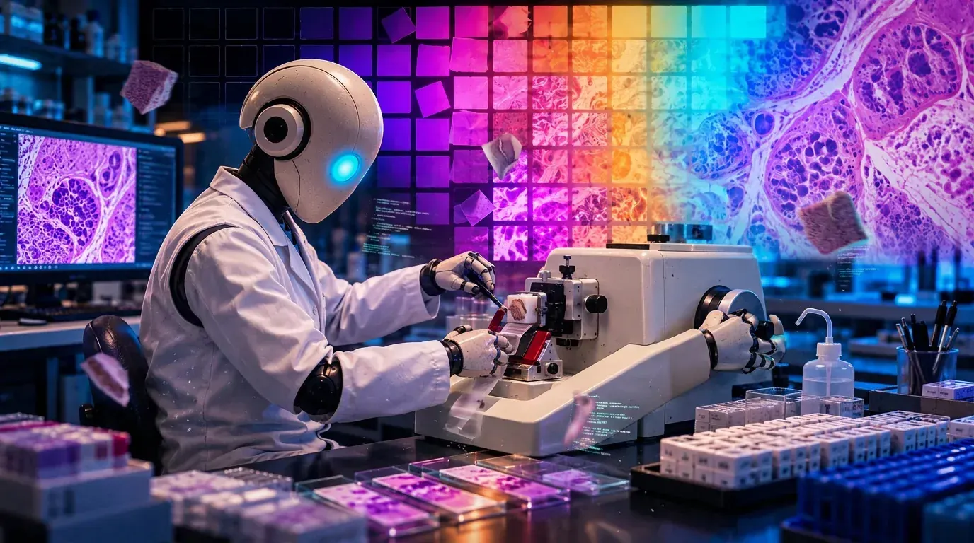
Artificial intelligence is a label for software that performs tasks people associate with human thinking. Recognizing a face. Translating a sentence. Sorting an email into junk. Finding a tumor in an image. The label covers many different techniques, and most of them work the same way underneath.
Something goes in. The software runs it through a long chain of arithmetic. Something comes out. What makes the arithmetic useful is a process called training. Developers feed the system thousands or millions of examples along with the correct answers, and the system adjusts its own internal numbers until its answers match the correct ones often enough. Those internal numbers, usually called weights, are all the system keeps. Nothing in there holds a concept of what prostate tissue is or why a biopsy was taken. There is a set of numbers that, given a new input, produces a new output.
A few terms come up often enough to be worth sorting out. Machine learning describes any system that derives its own rules from examples instead of following rules a person wrote. Deep learning is a subset that uses layered networks, and it is behind nearly every capability jump of the past fifteen years. A large language model is deep learning applied to enormous quantities of text, trained to predict the next piece of writing given what came before. ChatGPT, Claude, and Gemini are large language models wrapped in a chat window.
One distinction matters more than the rest. Narrow systems do one task or a small group of related tasks, and every product on the market today is narrow, including the chatbots. General intelligence, meaning software that could handle any mental task a person handles without being rebuilt for each one, does not exist. Most of the confusing claims in the news come from mixing those two up.
Much of the lab already runs on the older, simpler end of this family. A tissue processor reads a sensor, compares the reading to a setpoint, and corrects. Reagent management software tracks solution use and pulls a station when a limit trips. Printers pull identity from the LIS. None of it gets marketed as artificial intelligence, because anyone can point to the sensor, the controller, and the program.
Digital pathology is where the newer tools entered histology, and scanning came first. In April 2017 the FDA authorized the Philips IntelliSite Pathology Solution through the De Novo pathway for primary diagnostic use, the first whole slide imaging system cleared to let a pathologist sign out from an image instead of glass. The clearance covered formalin-fixed paraffin-embedded surgical pathology and excluded frozen sections, cytology, and non-FFPE hematopathology. Scanning itself involves several trained components: autofocus choosing a focal plane across a section that varies in thickness, tissue detection deciding where the section sits on the slide, and stitching assembling the individual fields into one image.
Software that reads the scanned image followed. Paige Prostate received De Novo authorization in September 2021, the first AI-based pathology product cleared for in vitro diagnostic use. It marks areas on a scanned prostate biopsy that are suspicious for carcinoma and presents them to the pathologist, who still makes and signs the diagnosis. The supporting study used sixteen pathologists reading 527 digitized biopsy images, and detection improved on the assisted reads.
Quantitative image analysis is probably the application most histotechs will encounter first, and it has been running in research and pharma for years, since work outside clinical diagnosis needs no clearance. These platforms segment tumor from stroma, identify and count nuclei, measure staining intensity, and apply a threshold across an entire section rather than across the few fields a person can hold in mind at once. Ki-67 indices, H-scores, PD-L1 tumor proportion scores, and immune cell densities in multiplex panels all come out as continuous numbers that another person can reproduce.
Quality tools sit alongside those. A scanner can flag out-of-focus regions, incomplete tissue detection, and low contrast before a slide reaches a pathologist's queue. Analysis software can exclude necrosis, pigment, and edge artifact from a scored area, or refuse to score a slide whose hematoxylin falls outside the range it was built for. Some platforms normalize color across slides to reduce the effect of staining differences between runs.
Operational applications get the least attention. Tracking systems predict turnaround and flag cases likely to miss it. Service software schedules maintenance from usage counts and error logs rather than a calendar. Workload models weigh case mix instead of counting blocks. Language models draft documentation, summarize validation data, and produce first passes at reports, with a person responsible for everything that goes out.
All of these tools share one limitation that belongs on the bench. The software receives whatever the slide gives it, and it has no way to ask whether the section was representative or whether the run was good.
Anyone who has spent a career around instruments already knows how to handle this. A processor gets trusted to run and gets checked anyway. A stainer gets a control on every run. Software that scores tissue earns the same treatment, and for the same reason.
Related Essays:
AI in Histotechnology: Promise in Research, Friction in the Clinic
Picks up where the primer ends: the same tools stall in the clinic on economics, not capability.
What Artificial Intelligence Actually Is
Further reading on the mechanism, the naming history, and where the same logic runs outside the laboratory.
Thin Sections covers the judgment work in histology that procedure manuals leave unnamed. Free, every other week.
Thin Sections is written by Jerry Clarin, HT (ASCP). The views expressed are his own and do not represent any employer.